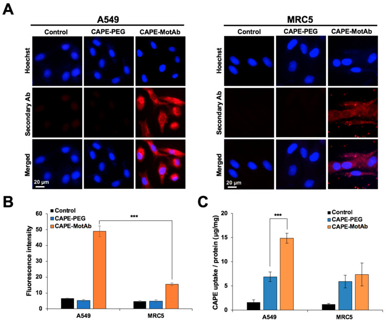
Figure 4.
Selective cellular uptake of CAPE-MotAb in cancer cells by targeting mortalin. (A) Fluorescence microscopy images of A549 and MRC5 cells treated with CAPE-PEG and CAPE-MotAb followed by staining with Alexa FluorTM 594-tagged secondary antibody. The nuclei were stained with Hoechst. Higher cellular uptake efficiency of CAPE-MotAb was observed in A549 cells. (B) Quantitation of mortalin expression from fluorescence images (mean ± SD, n = 3), *** p < 0.001 (Student’s t-test). (C) Quantitative analysis of cellular uptake of CAPE-PEG and CAPE-MotAb in A549 and MRC5 cells (mean ± SD, n = 3), *** p < 0.001 (Student’s t-test). CAPE-MotAb treated A549 cells exhibited higher CAPE accumulation as compared to MRC5 cells treated with the same nanoparticles. A549 and MRC5 cells for all above experiments were treated with an equivalent dose of CAPE (20 µg/mL) for 12 h.

