Figure 4.
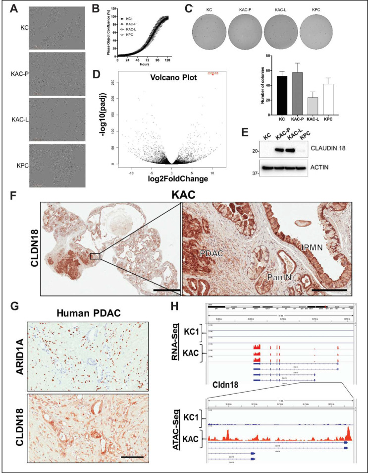
Characterization of autochthonous Arid1a-null PDAC cells from “KAC” mice. (A) In vitro monolayer cultures of “KC”, “KAC-P”, “KAC-L” and “KPC” cells revealed mesenchymal-like elongated morphology of “KAC” cells. Scale bar, 50 μm. (B,C) Assessment of growth in both monolayer (B) as well as soft-agar (C) showed no significant difference between “KAC” cells when compared to “KC” and “KPC”. Representative images of crystal violet stained colonies from 3 independent experiments. (D) Volcano plot of differentially expressed genes in “KAC-P” and “KC” cells, using RNA-Seq, showed Claudin 18 (CLDN18) as one of the top hit (n = 3). (E) Immunoblotting for mouse CLDN18 confirmed high expression levels in “KAC” PDAC cells compared to “KC” and “KPC”. Detailed information can be found at Figure S10. (F) IHC on pancreatic sections from “KAC” mice showed strong expression in epithelium of IPMN, PanIN, and PDAC, correlative with lack of ARID1A expression. Left, low magnification (2× objective) view of pancreatic section, Right, high magnification view (20× objective, Scale bar is 100 µm). (G) IHC on FFPE sections from human PDAC showed inverse relation between expression of ARID1A and CLDN18. Scale bar is 100 µm. (H) High transcript levels of Cldn18 in RNA-Seq corresponded to open chromatin at 5’ promoter region of Cldn18 gene by ATAC-Seq on “KC and “KAC-P” cells (n = 3).

