Figure 6.
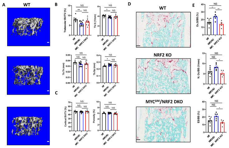
Myeloid-specific deletion of MYC decreases the osteoclast enhancing effect of NRF2 deficiency in vivo. (A–C) Micro-computed tomography (μCT) analysis of 12- to 13-week-old female WT, NRF2-deficient (NRF2 KO), and myeloid-specific MYC/NRF2-deficient (MYCΔM/NRF2 DKO) mice. (A) Representative μCT reconstructed images of the trabecular architecture of the distal femurs. Scale bar: 100 μm. (B) μCT measurements of the indicated parameters of the trabecular bone in the distal femurs. Bone volume/tissue volume ratio (BV/TV), trabecular numbers (Tb.N), trabecular thickness, (Tb.Th), and trabecular space (Tb.Sp) were computed using μCT analysis. (C) μCT measurements of the indicated parameters of the cortical bone in the midshaft of the femurs. BV/TV and porosity were computed using μCT analysis. Data are shown as mean mean ± standard deviation of at least seven mice per group. (D,E) Histomorphometric analysis of the trabecular bone in the distal femurs from 12- to 13-week-old female WT, NRF2 KO, and MYCΔM/NRF2 DKO mice. (D) Representative images showing the TRAP-positive, multinucleated osteoclasts (red-purple) in the coronal sections of the distal femur. Scale bar: 100 μm. (E) Histomorphometric analysis of the trabecular bone. Osteoclast surface area per bone surface (Oc.S/BS). Osteoclast number per bone surface (Oc.N/BS). Erosion over bone surface (ES/BS). All data are shown as mean ± s.e.m. of at least five mice per group. * p < 0.05 and ** p < 0.01 using one-way ANOVA in (B,E) except Tb.Th, which was analyzed using Kruskal‒Wallis test; NS, not significant.

