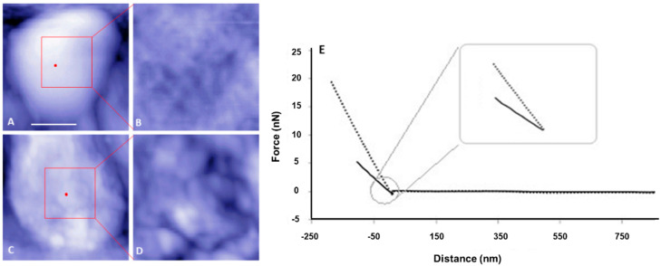
Figure 1.
Representative atomic force microscopy (AFM) topography images and corresponding force curves of live Rhizobium leguminosarum bv. viciae 3841 (wild type, top row) and 3845 (ctpA mutant, bottom row). Shown are low (A,C) and high (B,D) resolution AFM images of the live wild type (A,B) and ctpA mutant (C,D) rhizobia. Bar for A and C is 500 nm. Red dots on A, C indicate the approximate locations at the top center of the live cells, for collecting corresponding force approach curves (E) with solid (wild type) and dashed (ctpA mutant) lines, and subsequent creep experiments.

