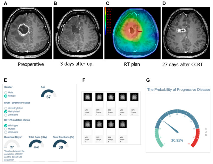
Figure 5.
Gadolinium-enhanced T1-weighted magnetic resonance (MR) images from a 45-year-old woman with glioblastoma and the clinical application of the machine learning model. (A) Pre-operative MR image showing an enhanced lesion. (B) No residual enhancing lesion in the cavity after the gross total resection. (C) Radiation therapy plan image showing the isodose line. (D) Enhancing lesion appeared in the resection cavity within the 80% isodose line after the completion of concurrent chemoradiation. (E) The screenshot of clinical information is given to the web platform. (F) Nine MR images are selected and uploaded to the platform. (G) Gauge figure representing the probability of progressive disease.

