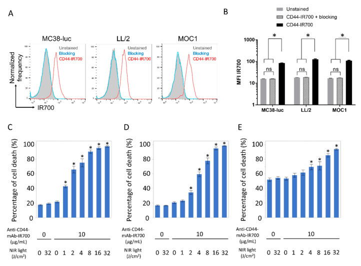
Figure 1.
Confirmation of CD44 expression as a target for NIR-PIT and evaluation of in vitro CD44-targeted NIR-PIT in MC38-luc, LL/2, and MOC1 cells. (A) Expression of cell-surface CD44 in MC38-luc, LL/2 and MOC1 cells was examined with flow cytometry. CD44-blocking antibody was added to some wells to validate specific staining. Representative histograms were shown. (B) Mean fluorescence intensity (MFI) of IR700 after labeling with anti-CD44-mAb-IR700 (n = 4, * p < 0.05, Tukey–Kramer test). (C–E) Membrane permeability as measured by PI staining, after labeling with anti-CD44-mAb-IR700 and treatment with NIR light. (C) MC38-luc; (D) LL/2; (E) MOC1 (n = 5, * p < 0.05, vs. untreated control; Mann–Whitney U test). ns, not significant.

