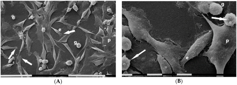
Figure 1.
Scanning electron microscopy of MDA-MB-231 cells cultured in 2D flask cultures. Cells only partially showed cell–cell contacts and mainly included “cobblestone” shaped flattened polygonal cells (p) and “squid” shaped elongated cells (e) with lamellipodia (arrows). Only very few isolated globular cells (g) were present. White bar = 100 µm (A). Three different cell phenotypes were distinguishable: flattened polygonal one (p), an elongated cell (e), and globular ones (g). Sparse microvilli were present on all cell surfaces and few microvesicles (arrows) were detectable on an elongated cell and globular ones. White bar = 10 µm (B).

