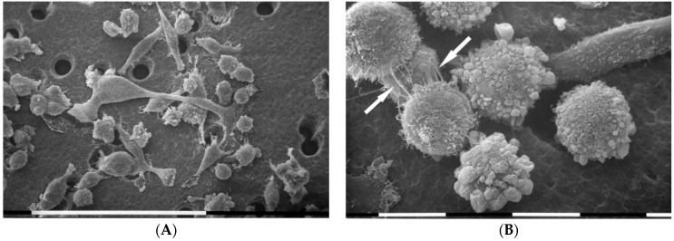
Figure 2.
Scanning electron microscopy of MDA-MB-231 cells cultured in Millipore filter. Two main cell phenotypes were observed: both globular cells and squid elongated ones showing lamellipodia and migrating through the Millipore filter holes. Microvilli and microvesicles mainly covering the globular cells were visible. White bar = 100 µm (A). Some globular cells show microvilli and cytoplasmic intercellular connections or tunneling nanotubes (TNTs) (arrows). Other globular cells exhibit many microvesicles. On the right and upside, an elongated cell showing only microvilli appeared to cross a Millipore filter hole. White bar = 10 µm (B).

