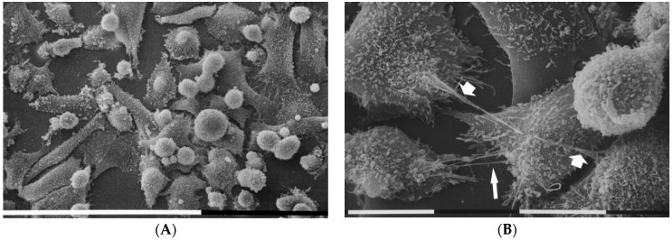
Figure 3.
Analysis of cell morphology in 3D cultures on Millipore filter covered by fibronectin (130 µg/mL) by scanning electron microscope. MDA-MB-231 cells cultured on fibronectin showed many “cobblestone” flattened polygonal cells and globular shaped ones, but few elongated ones. All cells exhibited both microvilli and many microvesicles. White bar = 100 µm (A). Flattened polygonal cells showing microvilli appeared to be connected by thin single TNTs (narrow arrow) and thicker ones (wide arrows) composed by single thin TNTs tightly bundled together. On the right, a globular cell exhibited microvesicles on the cytoplasmic surface. White bar = 10 µm (B).

