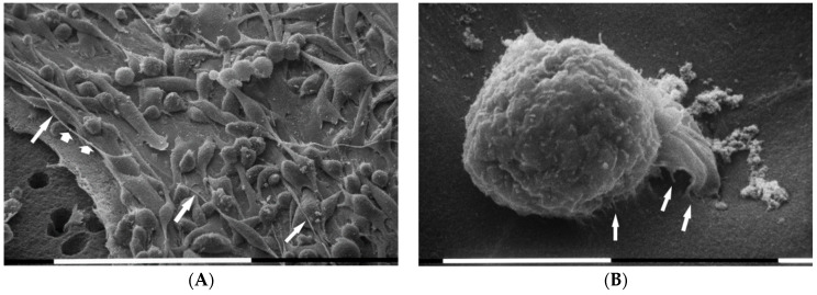
Figure 4.
Scanning electron microscopy (SEM) micrographs of 3D cultures on Millipore filter covered by concentrated Matrigel solution (3.0 µg/µL). MDA-MB-231 cells intimately appeared to be attached to the Matrigel layer. In cases where the Millipore filter was not covered by Matrigel (bottom left side of 4A), an elongated cell has almost completely crossed a non-Matrigel coated Millipore filter hole. Three phenotypes with microvilli and microvesicles were identified: the globular shaped cells, the flattened polygonal ones, and the elongated cells. In particular, some elongated cells developing long and thin filopodia (narrow arrows) sometimes containing microvesicles (wide arrows) display an evident fusiform shape. White bar = 100 µm (A). Short filopodia which morphologically could also resemble developing invadopodia (arrows) and a fully developed larger invadopodia (on the right) of a globular cell during the invasion of Matrigel are shown next to the substrate surface. Exosomes and microvescicles next to Matrigel surface can be seen on the left-upside and near the globular cell. White bar = 10 µm (B).

