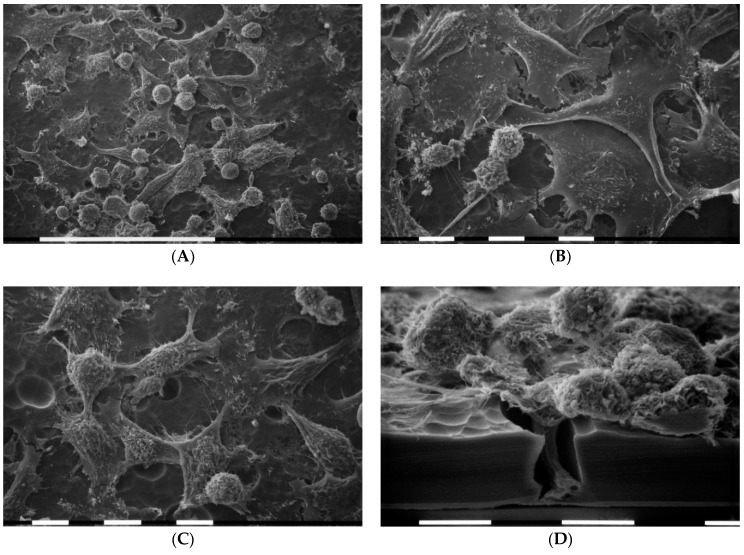
Figure 5.
Cell morphology of MDA-MB-231 breast cancer cells grown on Millipore filter covered by type I collagen (50 µg/mL). SEM analysis of 3D cultures showed that MDA-MB-231 cells include almost equally distributed and relatively isolated cells showing elongated, globular, and flattened polygonal shapes. Millipore filter was not completely covered by collagen so that its rough surface was discernable. White bar = 100 µm (A). Cells showed microvilli and microvesicles on their surface. Flattened polygonal cells mainly exhibited few superficial microvilli and showed some cell–cell contact (B), whereas microvesicles were mostly distributed on globular and elongated cells which display short and thin filopodia radially spread from the cells. White bar = 10 µm (B,C). Lateral view of a razorblade sectioned Millipore filter. Observe the wave rough porous Millipore surface: a collective invasion with a group of cells is taking place. The leader cell shows a “funnel” shape while invaginates and crosses a Millipore hole. White bar = 10 µm (D).

