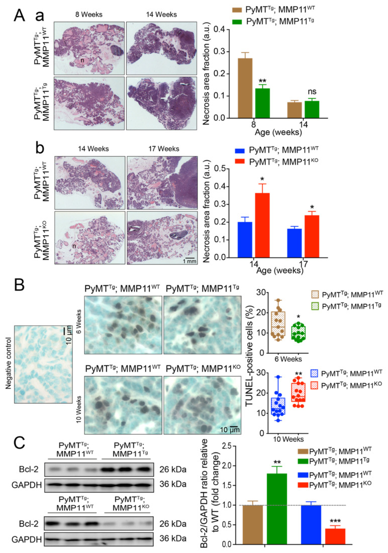
Figure 2.
MMP11 plays anti-necrosis and anti-apoptotic roles in early stage of tumor development. (A) (a) Hematoxylin and eosin (HE) staining shows decreased tumor necrosis area in 8-week-old PyMTTg; MMP11Tg mice but no change in necrosis area at 14-week-old mice as compared to in PyMTTg; MMP11WT age-matched mice; (b) 14- and 17-week-old PyMTTg; MMP11KO mice exhibit increased necrosis area as compared to control mice; (B) Apoptosis was measured by the TUNEL-assay apoptotic cells in tumors from 6-week-old PyMTTg; MMP11Tg mice as compared to age-matched controls (upper panel), whereas 10-week-old PyMTTg; MMP11KO mice exhibit a significant increase in apoptotic cell percentage as compared to control animals (lower panel); (C) Immunoblots of tumor protein extracts show an increase in the expression of the anti-apoptotic protein Bcl-2 in 6-week-old PyMTTg; MMP11Tg mice as compared to controls, while a decrease in Bcl-2 is observed in tumor protein extracts from PyMTTg; MMP11KO mice as compared to controls (left panel). Quantification of immunoblot signal density normalized to glyceraldehyde-3-phosphate dehydrogenase (GAPDH) protein levels relative to wild type is depicted in the right panel. N = 6–8 mice/group, data are expressed as mean ± SEM. * p < 0.05, ** p < 0.01, *** p < 0.001 (unpaired t-test).

