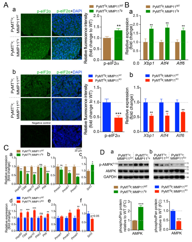
Figure 5.
MMP11 promotes tumor cellular endoplasmic reticulum (ER) stress response (UPRER) and alters mitochondrial UPR (UPRmt) in mammary gland tumors. (A) (a) Confocal images of tumor sections were stained using an anti-phosphorylated α subunit of eukaryotic translation initiation factor 2 (eIF2α) (p-eIF2α) antibody, from 12-week-old PyMTTg; MMP11Tg and control age-matched mice; (b) Confocal images of tumor sections stained with anti-p-eIF2α from 14-week-old PyMTTg; MMP11KO and control; a control staining without primary antibody is shown below; (a,b) relative image quantification are presented as histograms in the right; (B) (a) RT-qPCR analysis of genes implicated in UPRER in tumors from 6-week-old PyMTTg; MMP11Tg mice compared to tumors from control mice; (b) and in tumors from PyMTTg; MMP11KO mice compared to controls; (C) (a–c) Expression profile of genes implicated in the three arms of the UPRmt in tumor samples from 6-week-old PyMTTg; MMP11Tg mice compared to controls; (d–f) Expression profile of genes implicated in the three arms of the UPRmt in tumor samples from 10-week-old PyMTTg; MMP11KO mice compared to controls; (D) Western blot analysis of phosphorylated AMP-activated kinase (AMPK) (pAMPKT172) in tumors from (a) 6-week-old PyMTTg; MMP11Tg and (b) 10-week-old PyMTTg; MMP11KO mice, respectively, as compared to their controls. Quantification of the ratios of pAMPK/AMPK is presented below the blots, normalized to GAPDH expression. Data are presented as fold changes. N = 6–8 mice/group, data are presented as mean ± SEM, * p < 0.05, ** p < 0.01, *** p < 0.001 (unpaired t-test).

