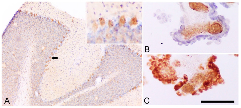
Figure 3.
TSPO is expressed by Purkinje and choroid plexus cells. (A) The immunoreactivity of rat cerebellum to TSPO (with cresyl violet counterstaining) underlines a positive staining in Purkinje cells (the black arrow indicates an example and insert), as demonstrated in mouse cerebellum [102]. (B) Immunoreactivity of human choroid plexus cells to CD31 (endothelial cells) with cresyl violet counterstaining (epithelial cells). (C) Immunoreactivity of human choroid plexus cells to TSPO underline both epithelial and endothelial cells positive for TSPO (as previously published in Reference [101]). Scale bar: 250 μm.

