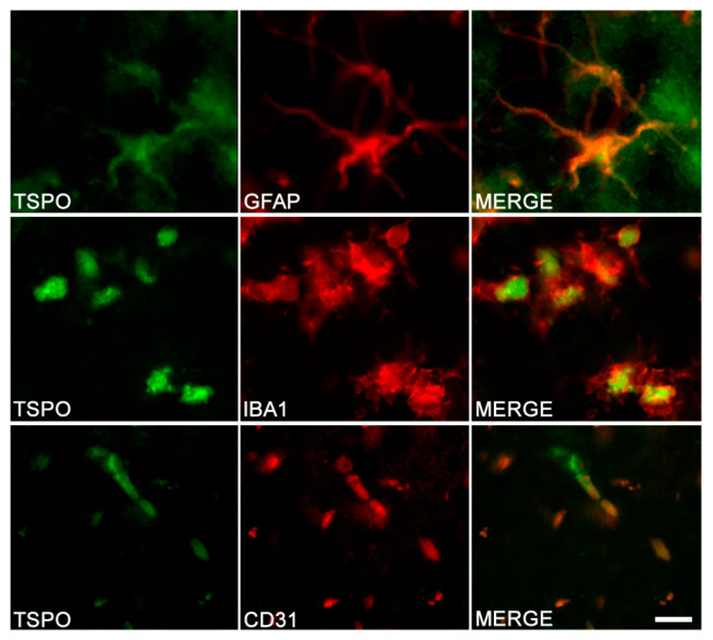
Figure 4.
TSPO in different cell types of the hippocampus in TgF344-AD rats. Double-immunostaining was performed to detect TSPO (left column) and specific marker of astrocytes (GFAP), microglia (IBA1) and endothelial cells (CD31). Merge images demonstrate the colocalization of TSPO with astrocytes, microglia and endothelial cells. Scale bar: 10 μm. Adapted from Reference [122].

