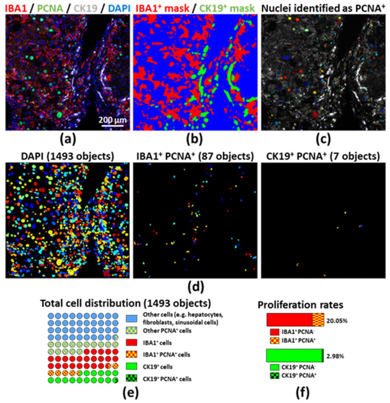
Figure 4.
(a) Multiplex immunostaining was performed on a FFPE section obtained from a DEN and CCl4 injected mouse (fibrosis–cancer model, compare to Figure 2); (b) Segmentation was performed using Ilastik; (c) Proliferating cell nuclear antigen (PCNA)-positive nuclei were identified and labeled on the original picture by using CellProfiler; (d) Single nuclei from every cell, monocyte/macrophages (IBA1+) or ductular cells (CK19+) were classified and counted using CellProfiler; (e) Segmented cells were numbered and the chart depicts the proportions of proliferating cells with the IBA1+, CK19+ or remaining cell compartments; (f) ratio of PCNA− versus PCNA+ monocytes/macrophages (upper panel) and ductular cells (lower panel). Additional masks used for this analysis shown in Figure S9.

