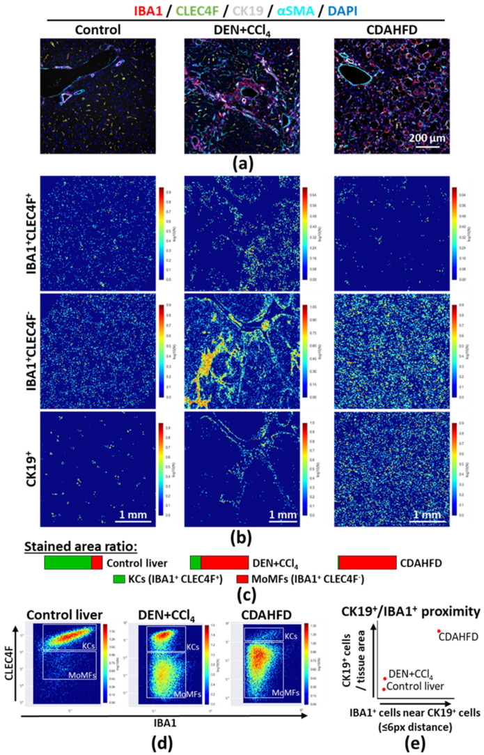
Figure 5.
(a) Multiplex immunostaining was performed on mouse FFPE liver sections to identify monocyte-derived macrophages (MoMFs, IBA1+CLEC4F−) and liver resident macrophages (i.e., Kupffer cells, KCs, IBA1+CLEC4F+) in healthy, DEN and CCl4 injected, choline-deficient, L-amino-acid-defined, high-fat diet (CDAHFD, 12 weeks) fed mice. Single-channel pictures are provided in Figure S10a–c; (b) MoMFs (IBA1+CLEC4F−), KCs (IBA1+CLEC4F+), and ductular cells (CK19+) were segmented using Ilastik, and spatial distributions were evaluated by using CellProfiler and CellProfiler Analyst; (c) Area covered by IBA1+CLEC4F− and IBA1+CLEC4F+ cells were measured in FIJI, using the masks generated in Ilastik; (d) Staining intensity for IBA1 and CLEC4F in individual stained cells was determined using CellProfiler; (e) CK19+ and IBA1+DAPI+ cells within 6 pixels from CK19+ cells were counted using CellProfiler, as described in Figure S12. Data represented as relative number of cells per total tissue area (arbitrary unit). Additional masks and enlargements provided in Figure S11a–f. Abbreviations: CDAHFD—choline-deficient; L-amino-acid-defined; high-fat diet; KCs—Kupffer cells; MoMFs—monocyte-derived macrophages.

