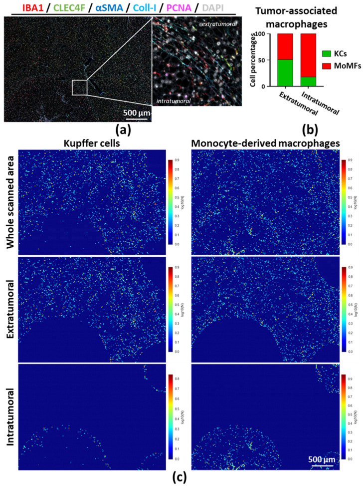
Figure 6.
(a) Multiplex immunostaining was performed on a liver FFPE section from a mouse that was injected with DEN and fed with a Western diet for 16 weeks (steatohepatitis–cancer model); (b) Tumor and extratumoral regions defined based on PCNA and collagen-1 stained areas and Kupffer cells (KCs, IBA1+CLEC4F+); monocyte-derived macrophages (MoMFs, IBA1+CLEC4F−) numbered as described above. Graph represents the relative contribution of KCs and MoMFs in the macrophage compartment of each region; (c) MoMFs and KCs segmented using Ilastik; and spatial distributions were evaluated by using CellProfiler and CellProfiler Analyst. Additional masks provided in Figure S13a,b. Abbreviations: KCs—Kupffer cells; MoMFs—monocyte-derived macrophages.

