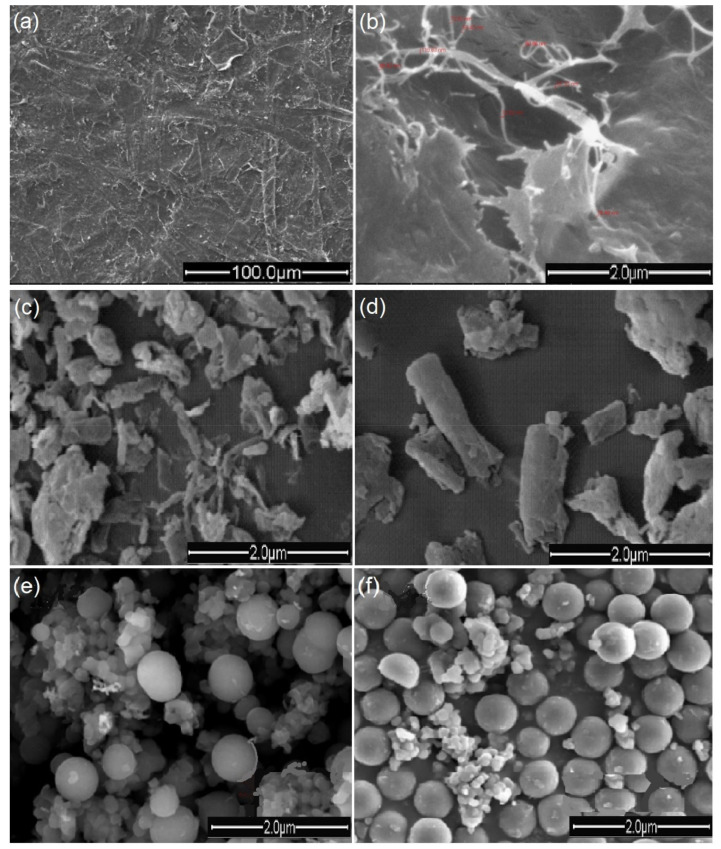
Figure 2.
Scanning electron microscope (SEM) images of different forms of microcellulose materials. (a,b) Surface of micro-fibrillated cellulose from the bleached cotton stalk [49], (c,d) the surface of micro-microcrystalline cellulose from the cotton stalk [50], (e,f) microcellulose particles of cotton, adapted [51].

