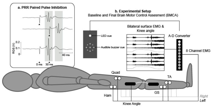
Figure 2.
(a) Example traces during posterior root reflex (PRR) testing; single pulses were delivered at 0 and 30 ms (upper traces) and as a pair with an interstimulus interval (ISI) of 30 ms (lower trace) to demonstrate paired pulse inhibition (arrows denote the time at which the stimulus was applied). (b) Experimental setup for baseline and final Brain Motor Control Assessments. Participants were placed in a supine position with bilateral electromyography (EMG) electrodes placed over the Quadriceps (Quad), Hamstring (Ham), Tibialis Anterior (TA) and Gastrocnemius (GS) muscles to record EMG and electro-goniometers were placed laterally across the knee joints to synchronously record knee joint range of motion.

