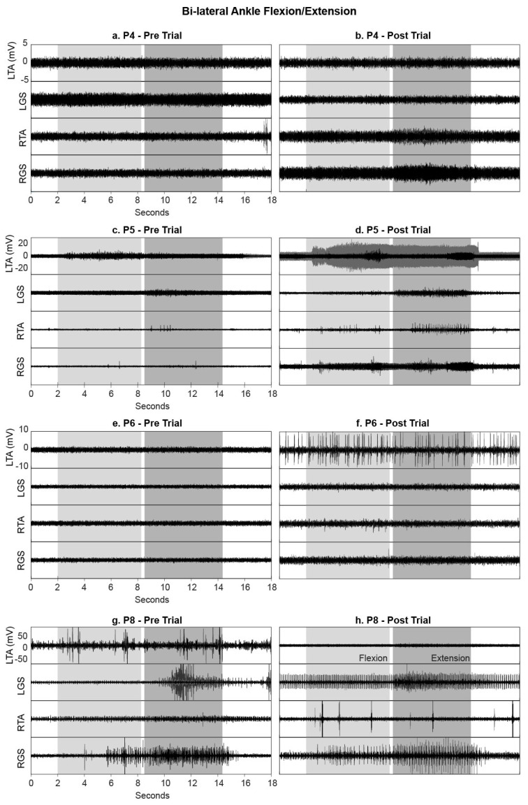
Figure 7.
EMG activity recorded from the left (L) and right (R) Tibialis Anterior (TA) and Gastrocnemius (GS) during ankle flexion (light grey) and extension (dark grey) movements without tSCS, before and after the intervention. Data are shown for (a,b) P4, (c,d) P5, (e,f) P6 (STIM group) and (g,h) P8 (NON-STIM group); each movement was repeated three times, and EMG data from all three movements are overlaid.

