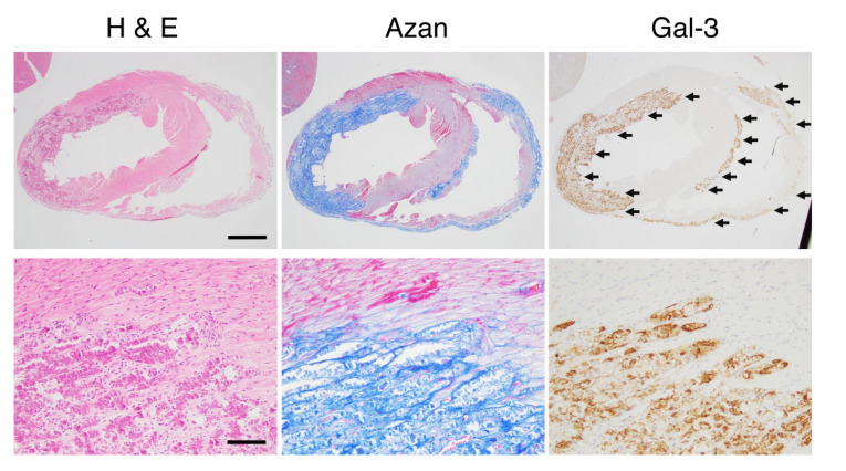
Figure 2.
The cardiac lesions of dilated cardiomyopathy in the late stage of δ-sarcoglycan (δ-SG) knockout (KO) mice. Microphotographs for hematoxylin and eosin (H&E) staining, Azan staining and immunohistochemistry of Gal-3 are shown. Scale bars in H&E = 1 mm in the upper panel and 100 μm in the lower panel. Gal-3 expression sites indicated by arrows are identical to the fibrotic areas detected as blue in azan staining.

