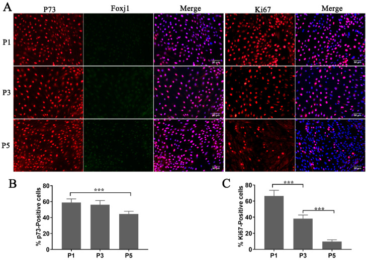
Figure 3.
Evaluation of the differentiation competency and proliferation rate in cryopreserved fallopian tube epithelium cells (FTECs) during expansion. (A) FTECs at different numbers of passages were stained with molecular markers for progenitor (p73, red), ciliated (Foxj1, green), and mitotic (Ki67, red) cells. They were counter-stained with DAPI (blue) to visualize nuclei for cell counting. Scale bars: 50 µm. (B,C) Quantitation of Ki67- and p73-positive cells (ANOVA test, n = 7). *** p < 0.001.

