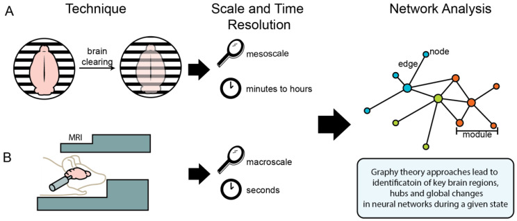
Figure 1.
Illustration of imaging techniques used for neural network analysis of functional and structural brain connectivity. (A) Single-cell whole-brain imaging techniques, such as iDISCO, allow for analysis of the whole brain at the mesoscale (i.e., with region and cell-specific resolution), with results representing neural activity across minutes to hours. (B) Magnetic resonance imaging (MRI) techniques allow for analysis of the brain in more generalized resolution (i.e., at the macroscale) in anesthetized or immobilized animals, with results representing neural activity across seconds. Data from both of these methods can be interpreted using graph theory approaches to identify key brain regions, hubs, and global changes in neural networks during a given state.

