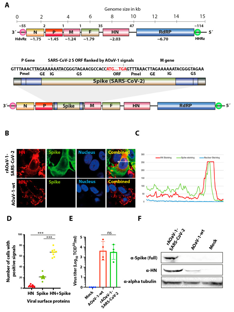
Figure 1.
Construction, rescue and characterization of rAOaV-1-SARS-CoV-2 vaccine candidate. (A) The full-length ORF for S gene of SARS-CoV-2 was over-hanged with required transcriptional signals (GE, GS, IG) and inserted in between P and M genes. The rough gene size is mentioned below each gene, the division of the genome across the length and number of nucleotides in intergenic region is displayed at the top of the schema of the rAOaV-1 genome. (B) Vero cells were infected with the rAOaV-1-wt or rAOaV-1-SARS-CoV-2 and stained for the expression of the HN (red) or S (green) proteins. The co-expression of both surface proteins is colored yellow in combined images and was marked with the arrow. (C) Quantitative co-expression profile is marked with arrow and shown in the line chart. (D) A total of nine microscopic fields were scanned for the presence of HN or S or both proteins. A significantly higher proportion of HN+S expressing cells were identified. (E) A comparable replication of rAOaV-1-wt or rAOaV-1-SARS-CoV-2 in Vero cells indicating the competitive replication of rAOaV-1 even after the expression of the foreign S gene. (F) Western blot analysis for the expression of the HN protein, indicating the active replication of the rAOaV-1 and S protein indicating the rAOaV-1-SARS-CoV-2, indicating the replication competence of the recombinant virus. Alpha tubulin was used as loading control. *** indicates statistically significant difference with p < 0.005, and ns indicates non-significant.

