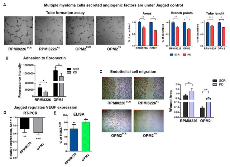
Figure 2.
MM cell-derived Jagged promotes angiogenesis: (A) Tube formation assay on HPAECs with conditioned media (CM) of HMCLsSCR or HCMLsKD. 4X magnification images are shown on the left. Graphs (on the right) show the percentage variation of areas and branch points and total tube length +/-SEM. (B) Adhesion to fibronectin of HPAECs treated with CM from HMCLsSCR and HMCLsKD and stained with Calcein-AM. The graph reports the intensity of the adherent fluorescent cells. (C) Motility of the HPAECs treated with CM of HMCLsSCR and HMCLsKD was assessed by wound healing assays. Up: Representative pictures at 4X magnification. Down: The graph shows the average open area of the wounds expressed in pixels. (D,E) Variation of vascular endothelial growth factor (VEGF) expression in HMCLsSCR and HMCLsKD assessed at the mRNA level (D) by qRT-PCR of the relative gene expression variation (normalized to GAPDH) calculated by the 2−ΔΔCt formula (data are expressed as the mean value ± SD) and at the protein levels (E) by ELISA on 48 h CM. Data are expressed as the amount of VEGF-A released by HMCLsKD normalized on VEGF-A expressed by HMCLsSCR. For each sample, the amount of VEGF-A (pg/mL) was normalized to the cell concentration. Statistical analyses were carried out by one-tailed t-tests; * is for p ≤ 0.05; ** is for p ≤ 0.01; *** is for p ≤ 0.001.

