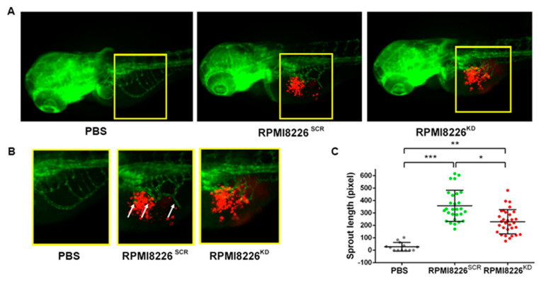
Figure 4.
Zebrafish embryo in vivo model to evaluate tumor-induced angiogenesis in relation to Jagged expression in HMCLs. (A) Representative images of Tg(fli1a:EGFP)y1 zebrafish embryos with GFP expressing vessels (green) and RPMI8226SCR or RPMI8226KD stained with CM-Dil fluorescent dye (red). Epifluorescence images were acquired with a Leica DM 5500B microscope equipped with a DC480 camera. (B) Inset of sprouting vessels from SIV in zebrafish embryos 24 hpi; white arrows indicate angiogenic sprouts. (C) Quantification of endothelial sprouts from the SIV plexus was performed in 24 h post-injection (hpi) zebrafish embryos injected with RPMI8226SCR (N = 24) and RPMI8226KD cells (N = 31) using ImageJ software (National Institutes of Health, USA). Statistical analysis was carried out by one-way ANOVA with Tukey post hoc tests of three independent experiments; * is for p ≤ 0.05; ** is for p ≤ 0.01; *** is for p ≤ 0.001.

