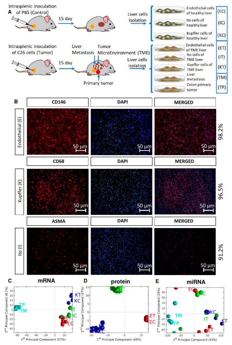
Figure 1.
Experimental design and global omics analysis. (A) Two groups of mice were inoculated with C26 murine colon cancer cells and with PBS (Control). After 15 days, the mice were perfused, and after PercollV R gradient centrifugation, three types of liver cells were collected, namely liver sinusoidal endothelial cells (E), Ito cells (I) and Kupffer cells (K), from control (C) and TME (T), and isolated to perform omics experiments (gene expression, miRNA expression microarrays and proteomics). TP and TM denote the CRC primary and tumor liver metastasis cells, respectively. (B) Cell purity was checked via immunochemistry using specific antibodies to detect endothelial (CD146), Kupffer (CD68), and Ito (ASMA) cells. (C–E) PCA plots of mRNA, protein and miRNA expression. Red, blue, and green symbols mark endothelial, Kupffer, and Ito cells, respectively. TME cells are depicted with dodecahedra, and controls with spheres. For each mRNA sample we used 4 biological replicates; for each miRNA sample 3 biological replicates, except 2 for ECs and KTs; for the proteomics data we used 9 biological replicates for ECs, 7 for ETs, 6 for ICs and ITs, and 5 for KCs and KTs.

