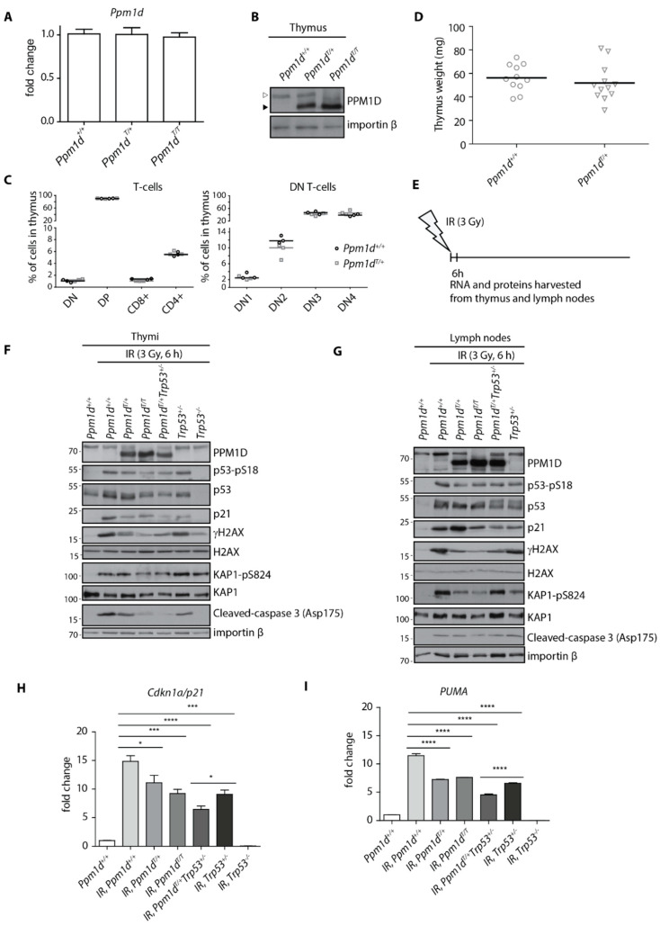
Figure 1.
Truncated PPM1D impairs DNA damage response in mouse thymus. Expression of PPM1D mRNA was analyzed by RT-qPCR in the thymi of Ppm1d+/+, Ppm1dT/+ and Ppm1dT/T mice and was normalized to GAPDH (n = 3) (A). Thymi from mice of indicated genotypes were lysed and proteins were separated by SDS-PAGE. Samples were probed with antibody against PPM1D and importin-β as a loading control. The empty and full arrowheads indicate the position of the full-length and the C-terminally truncated PPM1D, respectively. (B). Cells from thymi from Ppm1d+/+ and Ppm1dT/+ mice were analyzed by flow cytometry. Plotted are the counts of the indicated populations as follows: double-negative T-cells (DN and DN1, DN2, DN3, DN4), double-positive T-cells (DP), CD8-positive T-cells (CD8+) and CD4-positive T-cells (CD4+) (n = 3) (C). The median size of the thymus was determined in Ppm1d +/+ (n = 11) and Ppm1dT/+ (n = 12) mice (D). A scheme of the experimental setup in F-I. Mice were exposed or not to a low dose of IR (3 Gy), sacrificed after 6 h and thymi and lymph nodes were collected (E). Proteins isolated from thymi from mice of indicated genotypes exposed to mock or to IR were probed with the indicated antibodies by immunoblotting (F). Proteins isolated from inguinal lymph nodes from mice of indicated genotypes exposed to mock or to IR were probed with the indicated antibodies by immunoblotting (G). RNA isolated from thymi from mice in E was analyzed by RT-qPCR. The expression of CDKN1Ap21 mRNA was normalized to GAPDH. Statistical significance was evaluated by two-tailed t-test, error bars indicate SD, n = 5 (H). RNA isolated from thymi from mice in D was analyzed by RT-qPCR. The expression of PUMA mRNA was normalized to GAPDH. Statistical significance was evaluated by two-tailed t-test, error bars indicate SD, n = 5. * p <0.05; *** p < 0.0005; **** p < 0.0001 (I).

