
FIG 1.
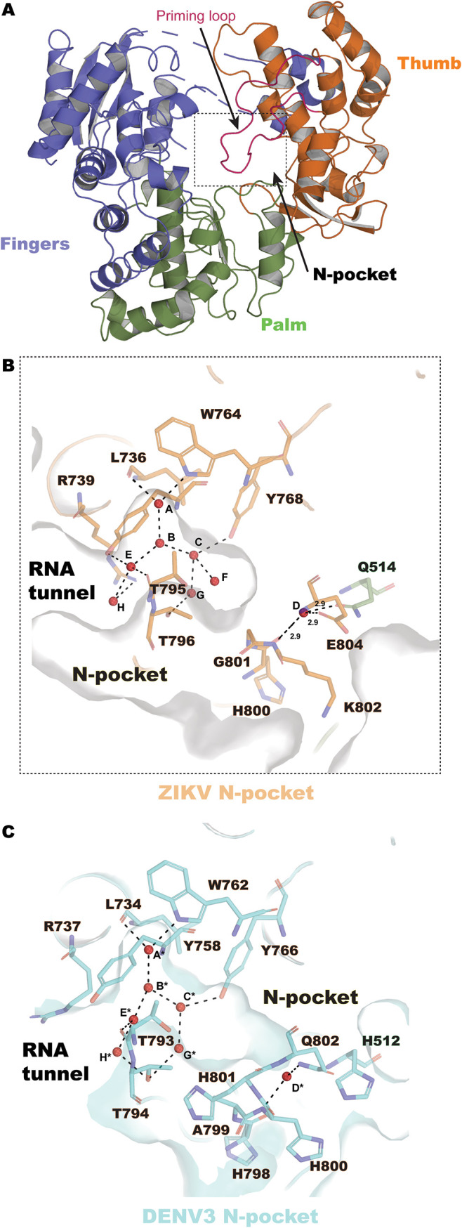
ZIKV RdRp structure at 1.4 Å and view of the N pocket. (A) Overall structure of ZIKV RdRp, represented as ribbons and colored according to subdomain (fingers in slate, residues 273 to 498 and 544 to 607; palm in green, residues 499 to 543 and 607 to 709; and thumb in orange, residues 710 to 887), and the priming loop region (residues 785 to 810) is shown in purple. (B) The hydrated N pocket of ZIKV RdRp is represented as a molecular surface and colored in gray. Eight water molecules (named A to H, red spheres) are inserted in the N pocket and are coordinated by residues from the thumb subdomain (represented as sticks, in orange). (C) The hydrated N pocket of DENV3 RdRp (PDB accession number 4HHJ) is represented as a molecular surface and is in cyan. Seven water molecules (named A* to H*, red spheres) are present in the N pocket and are coordinated by protein residues (represented as sticks).