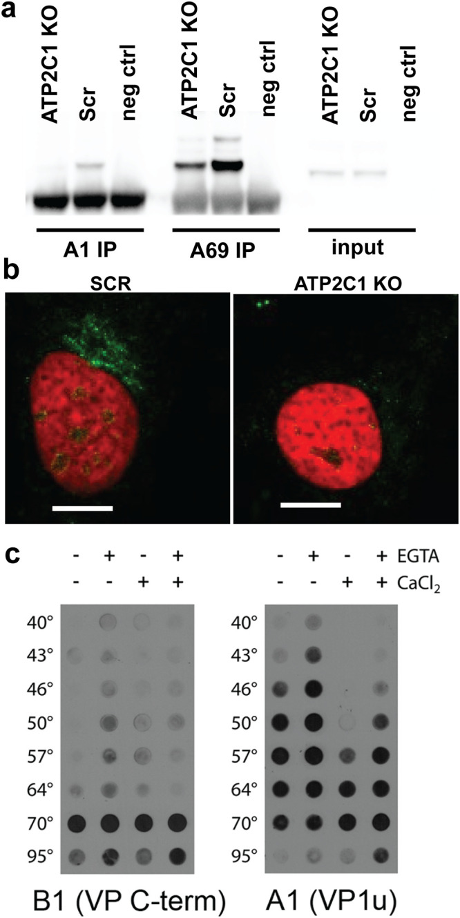
FIG 3.

ATP2C1 KO and calcium levels influence capsid conformational changes. (a) Immunoprecipitation of VP1 (A1) and VP1/VP2 (A69) of lysate from Scr and ATP2C1 KO Huh7 cells 18 h after transduction with AAV2 luciferase. (b) Confocal immunofluorescence microscopy of Scr and ATP2C1 KO Huh7 cells stained for externalized VP1/VP2 (A69). Bars, 10 μm. (c) Native dot blots with the B1 monoclonal antibody detecting the VP1 C-terminal domain and A1 monoclonal antibody detecting the thermally induced externalization of VP1 N termini from within the AAV capsid. Capsid stability assays were carried out using a thermal cycler with different AAV samples in the presence of the calcium chelator EGTA and/or extraneously added calcium ions.
