FIG 4.
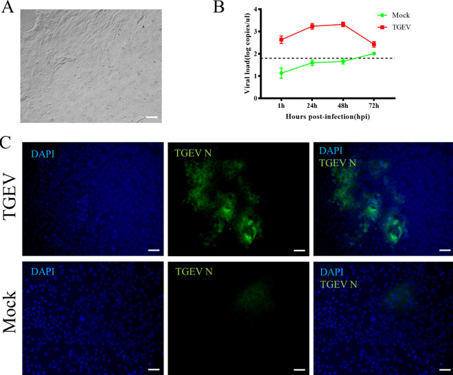
TGEV infection in porcine 2D intestinal organoids. (A) Organoids were dissociated and seeded on a Matrigel-precoated 24-well plate, and 2D monolayer organoids formed after 3 days in culture. (B) Then, 2D organoids were inoculated with TGEV, and samples were collected at the indicated time points for viral load detection by RT-qPCR. The dashed line represents the limit of detection. (C) TGEV-infected or mock-infected organoids were fixed at 48 hpi for staining with TGEV N protein (green). DAPI was used for nuclear staining. Images were obtained with a ZEISS Vert A1 microscope. Scale bars, 50 μm.

