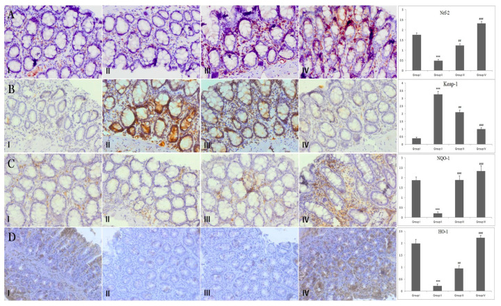
Figure 4.
Effect of piperine treatment on Nrf-2, Keap-1, NQO-1 and HO-1expression. Photomicrographs of colon sections depicting immunohistochemical analyses. Adjacent to photomicrographs are four panels which show quantitative evaluation of Nrf-2, Keap-1, NQO-1 and HO-1 expression immunostaining. Significant differences were indicated by *** p < 0.001 when compared with group I and (## p < 0.01), (### p < 0.001) when compared with group II (A) Brown color indicates specific immunostaining of Nrf-2, and light blue color indicates counter-staining by hematoxylin. The colonic section of DMH-administered group-II has decreased immunopositive staining of Nrf-2, as indicated by brown color, as compared to control group-I, while treatment of Piperine (30 and 60 mg/kg b. wt.) in groups-III and IV increased Nrf-2 compared to group II. Piperine significantly activated Nrf-2 in group III and IV, respectively, (## p < 0.01) and (### p < 0.001), when compared with DMH-administered group-II. (B) Photomicrographs of colon sections depicting immunohistochemical analyses; brown color indicates specific immunostaining of Keap-1 and light blue color indicates counter staining by hematoxylin. The colonic section of DMH-administered group-II has more Keap-1, as indicated by brown color, as compared to control group I, while treatment of piperine (30 and 60 mg/kg b. wt.) in groups III and IV reduced Keap-1 immunopositive as compared to group II. Piperine significantly suppressed Keap-1 in group III and IV, respectively, (## p < 0.01) and (### p < 0.001), when compared with DMH-administered group-II. (C) Photomicrographs of colon sections depicting immunohistochemical analyses; brown color indicates specific immunostaining of NQO-1 and light blue color indicates counter-staining by hematoxylin. The colonic section of DMH-administered group-II has decreased immunopositive staining of NQO-1, as indicated by brown color, as compared to control group I, while treatment of piperine (30 and 60 mg/kg b. wt.) in groups III and IV increased NQO-1 as compared to group II. Piperine significantly upregulated NQO-1 in group III and IV, respectively, (### p < 0.001) when compared with DMH-administered group-II. (D) Photomicrographs of colon sections depicting immunohistochemical analyses; brown color indicates specific immunostaining of HO-1 and light blue color indicates counter-staining by hematoxylin. The colonic section of DMH-administered group-II has decreased immunopositive staining of HO-1, as indicated by brown color, as compared to control group I while treatment of piperine (30 and 60 mg/kg b. wt.) in groups III and IV increased HO-1 as compared to group II. Piperine significantly upregulated HO-1 in group III and IV, respectively, (## p < 0.01) and (### p < 0.001), when compared with DMH-administered group-II. All images have original magnification of 40× (n = 10).

