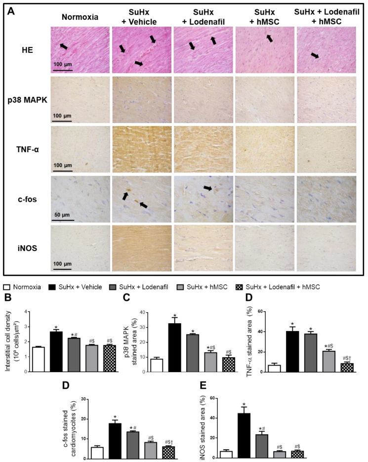
Figure 6.
Effects of lodenafil + hMSCs therapy on inflammatory markers in the RV from SuHx + PAH rats. (A) Representative images of histological and immunohistochemical stainings in RV fields. (B) Number of interstitial cells per μm2 in RV tissue area. (C) RV tissue area positively stained with p38 MAPK. (D) RV tissue area positively stained with TNF-α. (E) Number of cardiomyocytes positively stained with c-fos. (F) RV tissue area positively stained with iNOS. Data are expressed as mean ± SEM (n = 6). * p < 0.05 compared to normoxia group; # p < 0.05 compared to SuHx group treated with vehicle; $ p < 0.05 compared to SuHx group treated with lodenafil; † p < 0.05 compared to SuHx group treated with hMSCs. HE, hematoxylin-eosin; hMSCs, human mesenchymal stem cells; iNOS, inducible nitric oxide synthase; MAPK, mitogen-activated protein kinase; PAH, pulmonary arterial hypertension; RV, right ventricle; SuHx, SU5416/hypoxia; TNF-α, tumor necrosis factor alpha.

