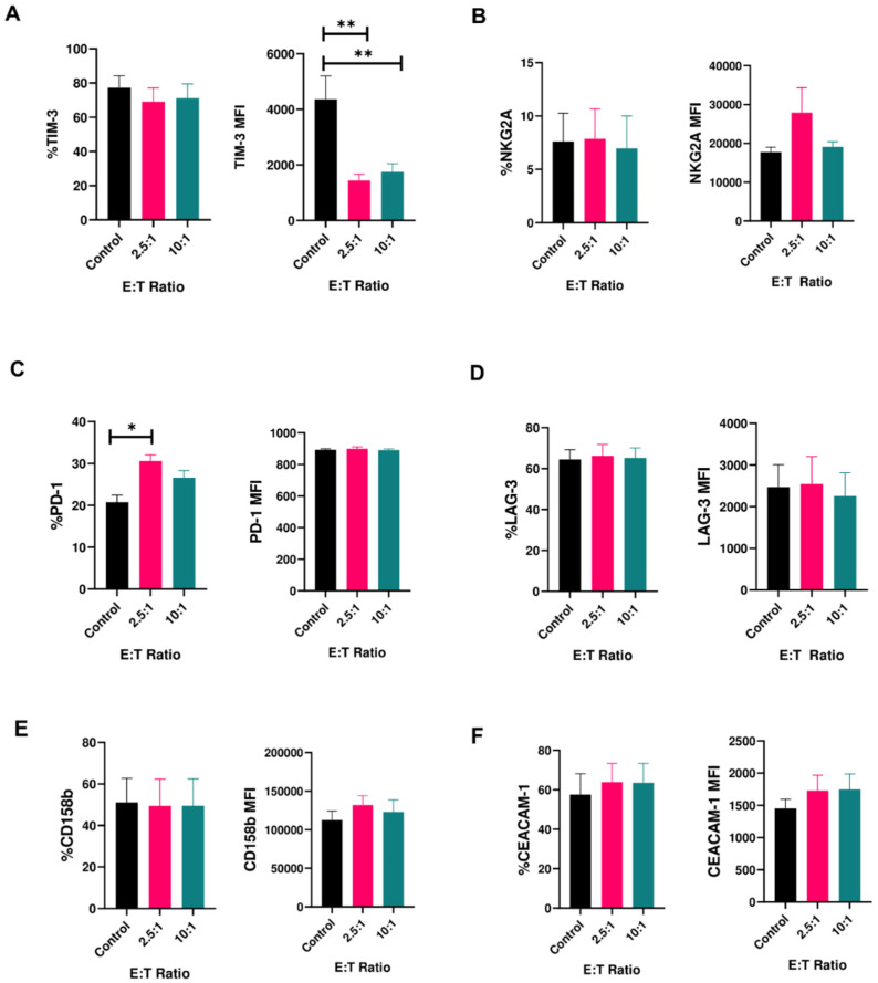
Figure 1.
Expression of inhibitory receptors on human NK cells in response to cancer cells (mean ± SEM). Percentage (left panels) and median fluorescence intensity (MFI) (right panels) of (A) T-cell immunoglobulin and mucin-containing domain (TIM-3); (B) NGK2A; (C) PD-1; (D) LAG-3; (E) CD158b; (F) CEACAM-1 on peripheral blood-derived human NK cells upon co-culture with U87MG cells for 4 h at effector:target (E:T) ratios of 2.5:1 and 10:1 (n = 6–9 samples). * p < 0.05, ** p < 0.01.

