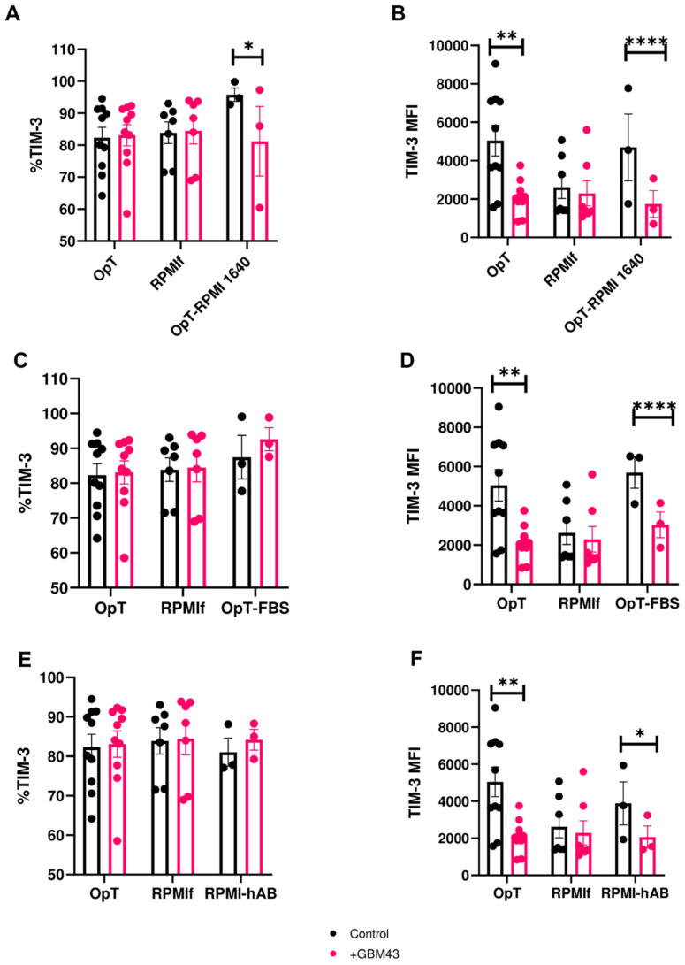
Figure 4.
Expression of TIM-3 on NK cells in response to cancer cells under varying media and serum compositions (mean ± SEM). Percentage and MFI of TIM-3 were measured on human NK cells after 4 h co-culture with GBM43 cells at an E:T ratio of 2.5:1 (n = 3–10 donors). (A,B) TIM-3 expression on NK cells expanded in OpT (n = 10), RPMIf (n = 7) or OpT with RPMI-1640 as the basal medium component (OpT-RPMI 1640, n = 3); (C,D) TIM-3 expression on NK cells expanded in OpT, RPMIf or OpT with fetal bovine serum FBS instead of human AB serum (n = 3); (E,F) TIM-3 expression on NK cells expanded in OpT, RPMIf, or RPMIf with human AB serum instead of FBS (n = 3). * p < 0.05, ** p < 0.01, **** p < 0.0001.

