Fig. 1.
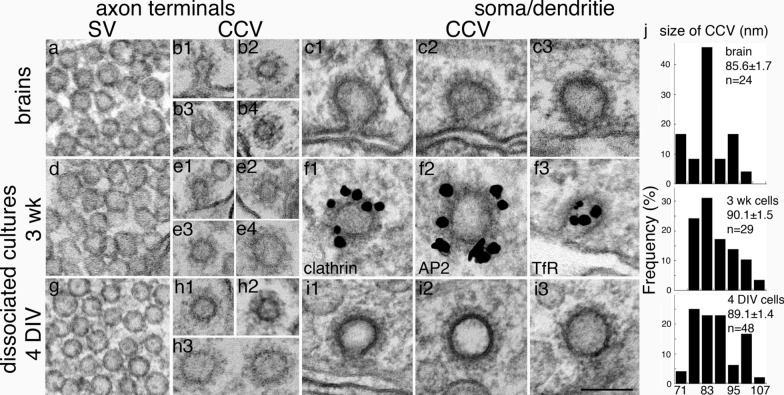
Clathrin-coated vesicles in axon and soma/dendrite are of different sizes. Images were sampled from perfusion-fixed mouse brains (top row: a–c), 3 wk-old (middle row: d–f) and 4 DIV (bottom row: g–i) dissociated rat hippocampal cultures. Synaptic vesicles (SV) were included as size references for CCV from the respective samples, and SVs from these three different groups of samples were of the same uniform size at ~ 40 nm in diameter (a, d, and g). CCVs in axon terminals from brains (b1–b4) were of the same size as SV. While the great majority of CCVs in axon terminals from dissociated cultures (e1-3; h1-2) were also of the same size as SV, some CCVs (e4; h3) were larger (~ 70 nm) than SV. In soma/dendrites (c, f, i), CCV were ~ 90 nm in diameter, much larger than those in the axon terminals. Immunogold labeling of 3 week-old cells illustrates that CCV in soma/dendrites labeled for clathrin (f1), AP2 (f2), and transferrin receptor (TfR, f3). j Histograms of size distribution of CCVs of soma/dendrites from brains (top panel), and dissociated neuronal cultures at 3 weeks (middle panel) and 4 days (bottom panel) in culture. The ranges of average diameter were similar (70–110 nm) among the three types of samples, and there was no statistical significance in mean values (ANOVA) or in median values (Wilcoxon test, median values at 85.8, 88.3, 88.3 nm, respectively). n = number of CCVs measured. Scale bar = 100 nm

