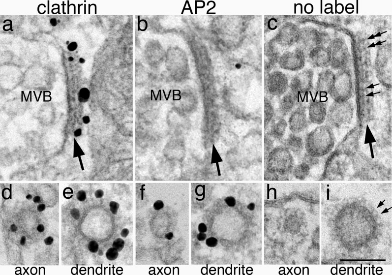
Fig. 4.
Multivesicular bodies (MVB, top row) in neurons contain a dark patch (large arrows) that label for clathrin (a) but not for AP2 (b). In contrast, in the lower row, clathrin-coated vesicles in axons (d, f) and dendrites (e, g) label for both clathrin (d, e) and AP2 (f, g). In samples fixed with glutaraldehyde for better structural preservation (no label, right column), a two layered arrangement with a uniform periodicity is visible (small arrows in c). The thickness of this patch is greater than those of the coated vesicles in axons (h) or in dendrites (i). MVBs in top row were sampled from neuronal soma (a) and dendrites (b, c). Scale bar = 100 nm

