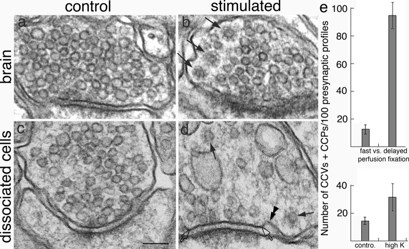
Fig. 5.
CCVs were uncommon in presynaptic terminals of fast perfusion-fixed brains (a), but became more abundant (arrows in b) upon a 5 min delay in perfusion fixation (b). Samples were from cerebral cortex of the mouse brain, and SVs were not noticeably depleted or dispersed in the delayed perfusion-fixed brains (b). Bar graphs in upper panel of e represent means of seven pairs of samples (data from Additional file 5A). In dissociated hippocampal cultures, upon depolarization with high K+, synaptic vesicles were typically dispersed and depleted (d). CCVs (arrows in d) were sometimes seen more frequently in high K+ (d) than in control samples (c). A CCP (double arrowheads in d) was seen adjacent to the active zone (between two open arrows) of the synapse. Bar graphs in lower panel of e represent means of four experiments (data from Additional file 5B). Scale bar = 100 nm

