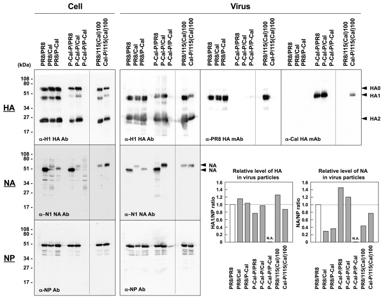
Figure 5.
Relative protein levels in RG viruses with chimeric HA and NA. MDCK-1 cells were infected with RG viruses at a MOI of 1.0 and were incubated at 34 °C for 1 d (2 days for P-Cal-P/P-Cal). The RG viruses in the culture media were concentrated through 20% sucrose cushions. The cell and virus samples were subjected to Western blotting with anti-H1 HA rabbit Ab, anti-PR8 HA mAb, anti-Cal HA mAb, anti-NA sheep Ab, and anti-NP rabbit Ab. Representative blots are shown. The band intensities in the virus samples proved by anti-H1 HA rabbit Ab, anti-NA sheep Ab, and anti-NP rabbit Ab were semi-quantified with ImageJ software. The levels of viral particles were normalized to the levels of NP in viral particle fractions. Relative levels of HA/NA in virus particles were evaluated as the ratio of HA/NA to NP. N.A.—not applicable.

