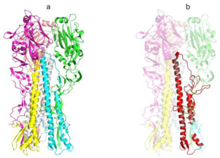
Figure 2.
The spatial structure of influenza virus A/Puerto Rico/8/1934(H1N1) HA protein and AgH1 antigen (extracellular portion). (a) The structure of the extracellular portion of influenza virus hemagglutinin protein trimer (PDB ID: 1RU7), elements of the secondary structure are shown with the ribbon diagram, individual chains have distinct colors and (b) the spatial structure model of artificial AgH1 antigen is shown with red color, the template HA structure is transparent. The picture was produced in PyMOL [45].

