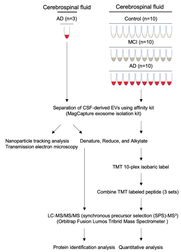
Figure 1.
Workflow used in proteomics analysis of CSF-derived EVs: EVs were separated from control, MCI, and AD CSF using the Affinity Capture method (MagCapture Exosome Isolation kit). The separated EVs were denatured, reduced and alkylated, followed by Lys-C and trypsin digestion, and labeled with a TMT 10-plex isobaric label kit for quantitative proteomics analysis. The non-labeled peptide (left) and combined TMT-labeled peptide (right) were analyzed by MS3 on an Orbitrap Fusion Lumos Tribrid Mass Spectrometer.

