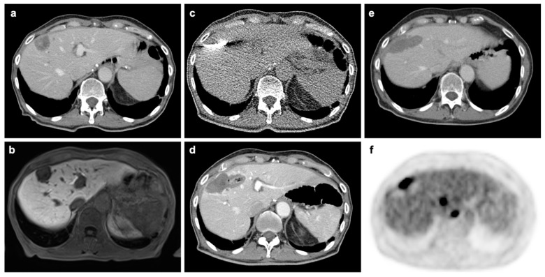
Figure 4.
Patient from our institution undergoing microwave ablation (MWA) for oligometastatic liver disease. (a). Pre-interventional CT in portal-venous phase shows contrast-enhancing metastasis in segment VIII. (b). Pre-interventional MRI in hepatocyte-specific phase. (c). Interventional imaging during MWA. (d). CT in portal-venous phase acquired immediately after intervention without evidence of complication or residual tumor. (e). Four-month follow-up F-18 FDG PET/CT without morphological evidence of recurrence in CT component. (f). PET-component acquired at same scan 4-month after MWA with tracer accumulation at the margins of the previous ablation in line with recurrence. Additional metastases without morphological evidence in the CT component can also be depicted.

