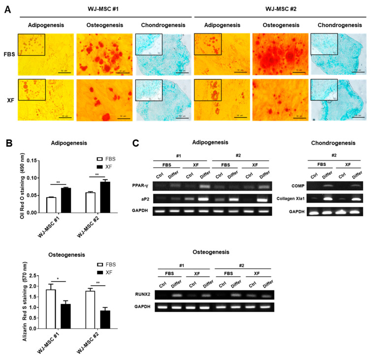
Figure 2.
Differentiation potential of WJ-MSCs cultured in FBS and XF conditions. WJ-MSCs cultured in FBS and XF conditions were cultured with adipogenic, osteogenic, chondrogenic differentiation medium for 21 days. (A) Light microscopic images show representative adipocyte, osteocyte, and chondrocyte. The lipid droplets were visualized using Oil Red O staining on adipogenic cells and calcium phosphate deposits were stained with Alizarin Red S. Glycosaminoglycans of the chondrogenic differentiated cells were stained with Alcian Blue. Representative images from at least three independent experiments are shown. (B) Quantification of the released dye of Oil Red O staining and Alizarin Red S staining were measured by a spectrophotometer. Data are represented as the means ± S.D. of triplicate experiments (* p < 0.05, ** p < 0.01). (C) Expressions of adipogenic (PPAR-γ, aP2), osteogenic (RUNX2), and chondrogenic (COMP, Collagen Xla1) genes were determined by RT-PCR with GAPDH as the reference gene.

