Figure 1.
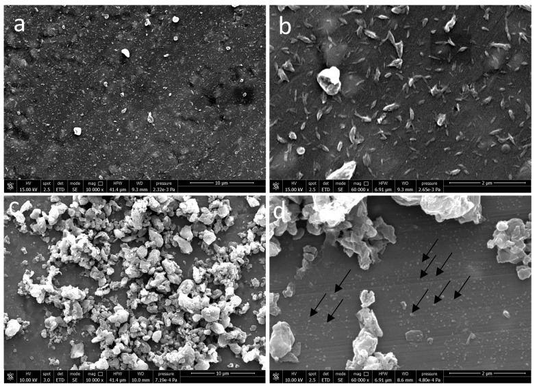
Scanning electron microscopy (SEM) micrographs of (a,b) electrosprayed CNs at 10,000× and 60,000× magnifications and (c,d) electrosprayed CN -NL assembled into micro-complexes loaded with (GA), i.e., (CLA) complexes, at 10,000× and 60,000× magnification. Arrows show subpopulation of CLA complexes with low size.

