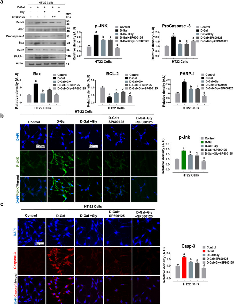
Fig. 6.
Glycine treatment reduced d-galactose-mediated elevated p-JNK-dependent neuroapoptosis in HT22 cells lines. a Representative western blot analysis of activated phosphorylated (p-JNK), procaspase-3, BCL2-associated X protein (Bax), Bcl-2 (B-cell lymphoma 2), and poly [ADP-ribose] polymerase 1 (PARP-1) proteins expression levels with or without JNK inhibitor (SP600125) in the HT22 cell line. The cropped bands were quantified using ImageJ software, and the differences are represented in the histogram. The density values are expressed in arbitrary units (A.U.) as the mean ± SEM for the respective indicated protein. An anti-β-actin antibody was used as a loading control. Number of experiments performed N = 3. b, c Immunofluorescence images of activated p-JNK (green) and caspase-3 (red) proteins along with their relative histograms after drug treatment with d-gal (100 mM), Gly (20 μg/μl), and SP600125 (20 μM) treatment in HT22 cell line for 24 h. The relative integrated density values are represented in arbitrary units (A.U) as the means (± S.E.M) for the respective indicated proteins. DAPI (blue) was used for nucleus staining. The data are expressed as the mean ± SEM. Magnification × 40. Scale bar; 50 μm. a Significantly different from the control group while bcd significantly different from the d-gal-treated groups. Significance: a, b, c, dP < 0.05

