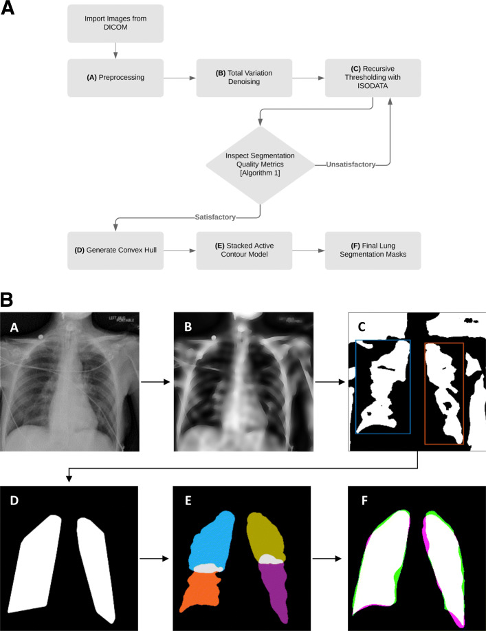
Fig. 1.
General outline of the proposed total-variation based active contour (TVAC) method. (a) An example source image containing a few wires from a patient diagnosed with acute hypoxic respiratory failure is shown. This image is normalized with contrast-limited adaptive histogram equalization (CLAHE) at this step. (b) Total variation denoising is used to diffuse wires while preserving edges of the lungs. (c) The denoised image is binarized with recursive thresholding and initial lung segments are extracted. (d) Convex hulls are generated from the extracted lung regions to enclose the lung fields and capture regions lost during binarization. (e) Lungs are partitioned into quadrants; each is individually processed with the stacked active contour model to better capture “difficult” regions such as the apex and costophrenic recess. (f) Final output of the lung segmentation algorithm. Green represents the ground truth, magenta shows the algorithm’s segmentation output, and white illustrates overlap of the two – indicating regions that are correctly segmented. This example has a Dice coefficient of 0.9407

