
Fig. 2.
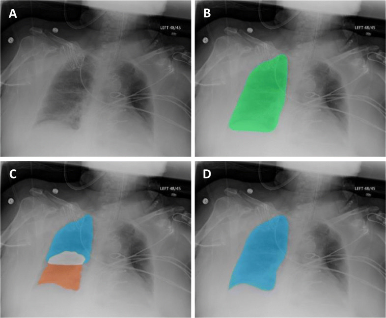
Segmentation with the stacked active contour model. (a) An example source image is shown for reference. (b) When the final segmentation mask is processed with a standard active contour model, areas of incorrect segmentation can be systematically observed – most commonly, at the right lung’s costophrenic recess and regions adjacent to the diaphragm. (c) Quadrant-based processing with a stacked active contour model shows better deformation and contouring to peripheral boundaries. (d) Final output for segmentation of the right lung after combining the upper and lower quadrants and applying a smoothing filter