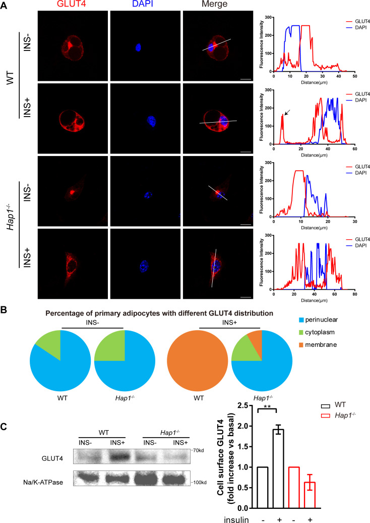
Figure 3.
Regulation of glucose transporter isoform 4 (GLUT4) translocation in adipose tissue by huntingtin-associatedprotein 1 (HAP1). (A) Immunofluorescence staining of GLUT4 (left) with cultured primary adipocytes from wild-type (WT) and Hap1-/- mice after 30 min insulin stimulation following 2-hour starvation and line-scan quantifications for the indicated lines drawn across the cells (right). The GLUT4 signal on the cell membrane is indicated by the black arrow. Scale bar, 20 µm. (B) The proportion of primary adipocytes with different GLUT4 immunofluorescence distribution at basal state and after 30 min insulin (INS) stimulation (n=24 for Hap1-/- cells and n=56 for WT cells) (χ2 test p<0.0001). (C) Detection of membrane-bound GLUT4 in primary Hap1-/- and WT adipocytes after 30 min insulin stimulation following 2-hour starvation by western blotting. Na/K-ATPase was used as a membrane loading control. Data are presented as mean±SEM from three independent experiments. **p<0.01 (unpaired two-tailed t tests). DAPI, 4′,6-diamidino-2-phenylindole.

