Figure 1.

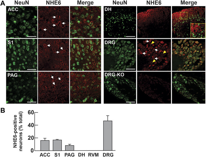
NHE6 is moderately expressed in pain centers in the CNS and highly expressed in sensory neurons. (A) Brain, spinal cord, and DRG tissues from 8-week-old WT mice stained for NeuN and NHE6. Representative images are shown (n = 3 mice, scale bar: 50 µm). White arrows and yellow arrowheads indicate cells positive and negative for NHE6, respectively. (B) Number of NHE6-positive neurons presented as a percentage of NeuN-positive neurons in each structure (ACC, n = 674/3288; S1, primary somatosensory cortex 1, n = 727/3556; PAG, n = 111/938; DH of the spinal cord, n = 6/701; RVM, rostral ventromedial medulla, n = 5/602; n = 474/989). ACC, anterior cingulate cortex; CNS, central nervous system; DH, dorsal horn; DRG, dorsal root ganglia; PAG, periaqueductal gray.
